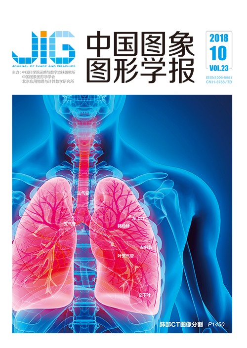
肺部CT图像中的解剖结构分割方法综述
摘 要
目的 高分辨率多层螺旋CT是临床医生研究肺部解剖结构功能、评估生理状态、检测和诊断病变的主要影像学工具。鉴于肺部各解剖结构间特殊的关联关系和图像成像缺陷、组织病变等干扰因素对分割效果的影响,学术界已在经典图像处理方法基础上针对CT图像中的肺部解剖结构分割进行了大量研究。方法 通过对相关领域有代表性或前沿性文献的归纳总结,系统性地梳理了现有肺组织、肺气管、肺血管、肺裂纹、肺叶或肺段等解剖结构CT图像分割方法的主要流程、方法理论、关键技术和优缺点,讨论了各解剖结构分割的参考数据获取、实验设计方法和结果评价指标。结果 分析了现有研究在结果精度和鲁棒性方面所面临的挑战性问题,以及基于分割结果在定位病变、定量测量、提取其他结构等方面展开的热点应用,特别详述了当前被重点关注的深度学习方法在本领域的工作进展,同时展望了本领域在分割理论方法和后续处理等步骤的发展趋势,并探索了如何在实践中根据分割结果发现新的临床生物标志。结论 快速精确地从CT图像中分割肺部各解剖结构可以获取清晰直观的3维可视化结构影像,展开解剖结构内部的定量参数测量或结构之间的关联关系分析能提供客观、有效的肺部组织疾病辅助诊断依据信息,可以大大减轻临床医生的阅片负担、提高工作效率,具有重要的理论研究意义和临床应用价值。
关键词
Review of anatomic segmentation methods in thoracic CT images
Bian Zijian1, Tan Wenjun2, Liu Jiren2, Zhao Dazhe2(1.School of Sino-Dutch Biomedical and Information Engineering, Northeastern University, Shenyang 110819, China;2.College of Computer Science and Engineering, Northeastern University, Shenyang 110819, China) Abstract
Objective Pulmonary disorder has high morbidity and mortality world-wide according to the reports by the World Health Organization.Some common pulmonary disorders include lung nodule and cancer,interstitial lung disease,chronic obstructive pulmonary disease,bronchiectasis,and pulmonary embolism.The disorders are typically characterized by long-term poor breath quality,irregular blood supply,and obstructive airflow and circulation.Pulmonary disorders bring not only enormous societal financial burden but also physical and mental suffering to patients.Thus,the recognition and comprehension of the disorders are widely considered the most basic and crucial medical tasks.High-resolution multi-slice computed tomography (CT) has received significant attention from pulmonologists and radiologists due to its allowance of investigating pulmonary anatomic function,assessing physiological conditions,and detecting and diagnosing pulmonary disorders.Hundreds of isotropic thin slices that are reconstructed real time from a single spiral CT scan can realize objective,repeatable,and non-invasive clinical inspections unlike traditional tools,especially in the early disease stage.However,the manual delineation,measurement,and evaluation of volumetric scans are extremely time-consuming and entail intensively laborious work load for clinicians.Therefore,the biomedical engineering community aims to develop semi-automated and fully automated segmentations in CT images through voxel-by-voxel labeling by using computer software to separate sub-divided pulmonary anatomic structures from one another.In the presence of unique inter-and intra-anatomy relationships and the impact of imaging defects,abnormalities,or other interference factors,classical image processing methods suffer from performance limitations.The anatomic CT visibility is attenuated and the morphology is deformed spatially and pathologically,which affect the segmentation results negatively.Several studies,usually incorporating traditional work and carefully defined processing rules,that focus on thoracic CT images have been employed.Method In this paper,a systematic review of anatomic segmentation methods for pulmonary tissues,airways,vasculatures,fissures,and lobes is presented by tracking and summarizing the representative or up-to-date published literature.In addition,several attractive practices and extractions of sub-divided or related structures,derived from segmented anatomic results,are also attached to corresponding anatomic subsections.They include the segmentation of adhesive,pleura nodular,and interstitial diseased lungs;the centerline extraction of airways and vasculatures;airway wall quantification and segmentation;pulmonary artery and vein separation;and pulmonary segment approximation.Moreover,for all the referred segmentation methods,the full implementation pipeline,background image processing methodology,and key techniques are presented to elaborate the result performance.Analogous methods are further classified on the basis of their designed frameworks or mathematical theories,and the merits and demerits of each method type are analyzed at the end of the classification content.In general,evaluating segmentations and comparing with other work are tedious for researchers mainly because of the difficulty in obtaining the ground truth.LOLA11,EXACT09,and VESSEL12 are three of the public and authoritative MICCAI grand challenges in chest image analysis.They are introduced for result comparisons in the directions of pulmonary tissue and lobe,airway,and vasculature.The complete procedure renders it difficult for the owners to construct their reference repository and quantify the submission performance.The reported standard generation approaches to other anatomic-based applications are illuminated in parallel.The evaluated indices contain the anatomic boundary alignment and volume overlap and the trade-off between true and false positive detection.Subsequently,the experimental validation approaches are explained based on the reference standard and indices.The qualitative and quantitative results of different methods are shown specifically with the description of the test datasets.Result On the basis of the individual anatomic topic,the existing challenges of state-of-the-art studies are presented in detail,highlighting the accuracy performance in true positive detection and false positive removal and the robustness performance against the diversity of CT scanners,imaging protocols,and the appearance of various abnormalities.A set of practical problems,such as lesion location,qualitative and quantitative anatomic measurement,and sequential component segmentation,are also discussed in the paper.Besides deep learning algorithms,convolutional neural network-based algorithms have rapidly become a preference of medical imaging institutions.At present,two mature chest CT imaging fields exist,namely,nodule detection and malignancy prediction in lung cancer screening and interstitial lung disease type classification.The major deep learning-based efforts are surveyed in terms of their contribution to pulmonary tissue bounding box localization and pulmonary airway segmentation and leakage removal,most of which were proposed in the recent two years.The improvements are compared with previous methods.In terms of the frontier requirements from scientific groups,industrial units,and pulmonology domains,future work trends and open issues are listed pertinent to methodology and post-processing steps along with the applications,such as the identification of pulmonary lesion sites and the subsequent task of anatomic segmentation.The parameters of anatomic measurement are of vital importance in characterizing the progress and severity of pulmonary disorders.Thus,several possible innovative points to achieve these biomarkers are also recommended.Conclusion Theoretical studies and clinical practices will benefit from implementing accurate,fast,and robust pulmonary anatomic segmentation from large amounts of CT images.The transfer or modification of successful pulmonary segmentation methodologies will facilitate the segmentation of other tissues,organs,and multi-modality images.Inter-and intra-structure measurements and the relationship mapping information obtained,ranging from global to local analyses,can provide objective and effective evidence for computer-aided pulmonary disease detection and diagnosis.The 2D transversal or 3D visualization of these points can present intuitive,legible,and proportional views of the anatomic structures with the help of volume rendering techniques and grayscale Dicom slices overlaid by chromatic tissue marks.The contribution of these aspects reduces the labor requirement from pulmonologists and radiologists,thereby increasing their efficiency significantly.Although deep learning algorithms are relatively immature and require improvement in terms of segmentation time cost and refinement steps,they have considerable potential and are worth studying for pulmonary segmentation areas,which are predicted to dominate the field in the coming years.
Keywords
CT images pulmonary segmentation pulmonary airway segmentation pulmonary vasculature segmentation pulmonary lobe segmentation
|


 中国图象图形学报 │ 京ICP备05080539号-4 │ 本系统由